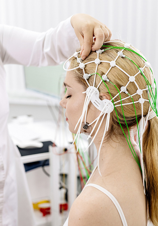
Electroencephalogram (EEG)​

An electroencephalogram (EEG) is a diagnostic test that measures electrical activity in the brain. This non-invasive procedure records brain wave patterns using small electrodes attached to the scalp.

What is an Electroencephalogram (EEG)?

There are two types of EEGs:

  1. Normal / Standard EEG (20 – 40 minutes)
  2. Long-Term / Full-Form (Video) EEG (Several hours / days)

These both record the electrical impulses produced by brain cells (neurons) through small electrodes placed on the scalp. These impulses are displayed as wave patterns on a computer screen or paper, allowing healthcare professionals to analyse the brain’s electrical activity.

An EEG is painless and safe, and it provides valuable information about the functional state of the brain.

Normal / Standard EEG

A normal EEG is a standard test conducted in a controlled environment, typically lasting 20 to 40 minutes. It evaluates baseline brain activity and helps detect any abnormal patterns that might indicate a neurological issue.

  • Epilepsy Diagnosis: Identifying abnormal electrical discharges that signal seizures.
  • Sleep Disorders: Assessing sleep-related abnormalities such as narcolepsy or sleep apnoea.
  • Head Injuries: Evaluating brain function after trauma.
  • Unexplained Symptoms: Investigating causes of fainting, confusion, or memory issues.

During the test, electrodes are placed on the patient’s scalp using a special adhesive or cap. The patient is asked to relax, with eyes closed, while performing simple tasks like deep breathing or looking at flashing lights to stimulate brain activity. The procedure is painless and carries no risks.

A neurologist analyses the recorded brain waves for abnormalities. Typical findings include rhythmic patterns in an awake state and slower waves during rest or sleep. Deviations from normal patterns may warrant further investigation.

Long-Term / Full-Form (Video) EEG

A long-term EEG, sometimes called video EEG monitoring, involves continuous recording over an extended period – ranging from several hours to several days. This test is ideal for capturing infrequent events like seizures or other episodic neurological symptoms that might be missed in a short EEG session.

  • Epilepsy Monitoring: Differentiating seizure types and localising their origin.
  • Unexplained Events: Evaluating recurring symptoms such as blackouts or muscle spasms.
  • Surgical Planning: Mapping brain activity before epilepsy surgery.

The setup is similar to a normal EEG, but the patient wears the electrodes for a prolonged duration, sometimes with a portable device. Video monitoring often accompanies the EEG to correlate brain activity with physical behaviour or symptoms. In an inpatient setting, patients stay in a monitored room with access to medical staff.

  • Captures rare or sporadic events.
  • Provides comprehensive data on brain function over time.
  • Enhances diagnostic accuracy for complex cases.

How is an EEG Performed?

The EEG procedure is straightforward and typically takes 30-minutes to an hour, although longer recordings may be required in some cases (ie: long-term EEG). The steps involved include:

  1. Preparation: The patient is asked to avoid caffeine and to wash their hair before the procedure, as this helps ensure good electrode contact with the scalp. No specific fasting or medication adjustments are usually required, but the patient should inform the technician of any medications they are taking.
  2. Electrode Placement: The technician attaches around 20 small metal discs (electrodes) to the scalp using a special adhesive or a cap fitted with electrodes. The electrodes are connected to an EEG machine by wires. A small amount of gel is applied to the scalp to improve the quality of the recording.
  3. Recording: The patient is asked to sit or lie down in a comfortable position and to relax. They may be asked to perform certain tasks during the recording, such as opening and closing their eyes, taking deep breaths, or looking at flashing lights. These activities can help provoke brain activity that may not be evident during rest.
  4. Monitoring: The EEG machine records the brain’s electrical activity, displaying it as a series of wavy lines on a monitor. Any abnormalities in these patterns can indicate issues with brain function.
  5. Completion: After the recording is complete, the electrodes are removed, and the patient can return to their normal activities. The results are analysed by a neurologist, who will interpret the wave patterns and identify any abnormalities.

Conditions Assessed with EEG at Canberra Neurology

EEG is used to diagnose and monitor a variety of neurological conditions, particularly those that involve abnormal brain activity. Some of the primary conditions include:

Overview:

  • Epilepsy is a chronic neurological disorder characterised by recurrent seizures, which are caused by sudden bursts of electrical activity in the brain. EEG is the primary tool for diagnosing epilepsy, as it can detect abnormal electrical patterns associated with seizures.

Symptoms:

  • Seizures, which can range from brief lapses in awareness (absence seizures) to full-body convulsions (tonic-clonic seizures)
  • Temporary confusion or altered consciousness
  • Uncontrolled jerking movements of the arms and legs
  • Sudden staring spells or brief episodes of unresponsiveness
  • Sensory disturbances, such as tingling, hallucinations, or unusual tastes or smells

Treatment Options:

  • Anti-Epileptic Drugs (AEDs): Medications are used to control seizures.
  • Surgery: In cases where medication is ineffective, surgery may be an option to remove the area of the brain where seizures originate.
  • Vagus Nerve Stimulation (VNS) and Responsive Neurostimulation (RNS): Devices that help control seizures in drug-resistant epilepsy.
  • Dietary Therapy: The ketogenic diet may be effective in reducing seizures in some individuals, particularly children.

Overview:

  • Encephalopathy refers to a broad category of brain disorders that affect brain function and structure. EEG can help diagnose the underlying cause of encephalopathy and assess the severity of the condition. It is often used in cases of altered mental status, confusion, or unexplained neurological symptoms.

Symptoms:

  • Confusion and disorientation
  • Memory problems
  • Difficulty speaking or understanding language
  • Changes in personality or behaviour
  • Drowsiness or lethargy
  • Seizures (in severe cases)

Treatment Options:

  • Treating the Underlying Cause: This may include antibiotics for infections, removing toxins, or managing metabolic imbalances.
  • Supportive Care: Ensuring proper hydration, nutrition, and oxygenation.
  • Medications: Anticonvulsants for seizure control and medications to manage symptoms such as agitation or anxiety.

An electroencephalogram (EEG) is a valuable tool in the diagnosis and management of various neurological conditions.

It provides crucial insights into brain function, helping clinicians diagnose conditions like epilepsy, encephalopathy, and a number of other disorders. If you or someone you know is experiencing symptoms suggestive of a neurological issue, such as seizures, altered mental states, or unexplained behavioural changes, it is important to seek medical advice.

A neurologist can determine whether an EEG is necessary and develop a comprehensive treatment plan tailored to the individual’s needs.

Helping to Restore Your Quality of Life

We are experts in the diagnosis and treatment of a range of neurological conditions. We offer the latest evidence-based medicine with the goal of helping you get back to doing what you enjoy.

Cutting-Edge Diagnostic & Therapeutic Neurology​

We offer the latest evidence-based diagnostic and therapeutic neurological care, spanning the fields of neurophysiology, electroencephalogram (EEG), and botox treatment.

Get in touch to make a booking.